Like many eye care practices, cataract surgery has undergone several developments over the years, continuously adapting to meet the various healthcare needs of today’s patients. To illustrate how the practice has evolved, we present a timeline showcasing the progression of how cataract surgery is done, from its earliest methods to the advanced techniques leveraged today by eye specialists at a leading Singapore eye clinic.
How Did Cataract Surgery Come to Be?
There is no concrete evidence as to where cataract removal surgery originated. However, an ophthalmic historian, Julius Hirschberg, once noted, “At the present time, it is impossible to answer this important historical question as to which nation (or even which man) first performed a cataract operation.” He also mentioned that there is no confirmation whether or not Indians invented the procedure.
However, this only implies that cataract surgery likely originated in one specific region and then spread globally. Some scholars believe this because there are striking similarities in the description of cataract surgeries from both Eastern and Western traditions, suggesting a single origin in the Old World.
Timeline of the Different Types of Cataract Surgery Techniques & Solutions
The history of cataract surgery is marked by several techniques. The types of cataract surgeries include:
1. Couching (500 – 401 BC)
Couching is the oldest known method of cataract surgery, believed to have been practised as early as the 5th century BC. This comes from the French word “coucher,” which means “put to bed.” This procedure involves using a sharp needle to pierce the eye close to the limbus until the practitioner can dislodge the cataract in the vitreous chamber and out of the visual axis.
While this method could potentially restore some level of vision, it was fraught with complications, such as infection and total vision loss, and the patient often remained at high risk of developing secondary cataracts.
2. Extracapsular Cataract Extraction (600 BC)
While couching was the predominant type of cataract surgery in the 18th century, the method of extracapsular cataract extraction also has historical roots dating back to 600 BC. Ancient texts credit the Indian surgeon Sushruta with the early practice of this technique, though it was French surgeon Jacques Daviel who experimented with the first documented true extraction.
This technique required a substantial corneal incision of more than 10mm. From this, the surgeon would then puncture the lens capsule, express the nucleus, and then remove the lens cortex through curettage.
However, extracapsular extraction was not without substantial risks. Patients often faced post-operative complications such as poor wound healing, retained lens fragments, posterior capsular opacification, and infection.
3. Intracapsular Cataract Extraction (1753)
Intracapsular cataract extraction was introduced by London surgeon Samuel Sharp. This procedure involves removing both the opacified lens and its surrounding capsule as a single unit. The method typically entails dissolving the zonular fibres that hold the lens capsule in place, followed by the extraction of the lens-capsule complex through a substantial limbal incision.
However, this technique came with significant risks, as the removal of the lens capsule often leads to complications, such as vitreous prolapse and potential retinal detachments. Additionally, because the procedure requires a large incision, patients tend to face longer recovery times and an increased risk of infection.

4. Intraocular Lenses (1949)
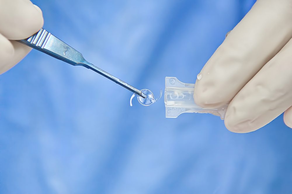
The concept of replacing the cloudy natural lens with an artificial intraocular lens was first implanted by Sir Harold Ridley in 1949. The first intraocular lens implant used by Ridley was made of polymethyl methacrylate (PMMA), also known as acrylic glass. During his time, the use of intraocular lenses garnered little support since there were considerable post-operative complications, like uveitis, glaucoma, and even the dislocation of implanted lenses.
However, this procedure changed the landscape of cataract surgery, facilitating the restoration of vision without the need for thick glasses or special contacts. Over time, the materials and designs of intraocular lenses have changed, with options that can be utilised for today’s commonly performed cataract removal approach, which is phacoemulsification.
5. Phacoemulsification (1967)
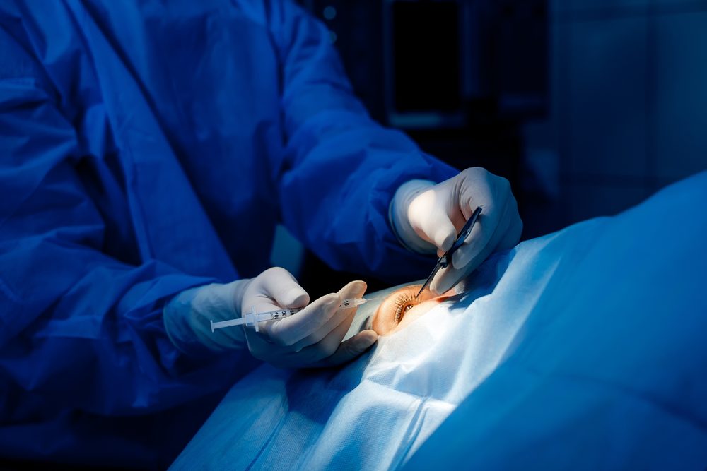
Introduced in the late 1960s by Dr. Charles Kelman, phacoemulsification was developed to potentially minimise downtimes and the side effects of modern cataract removal. This procedure uses ultrasonic vibrations to emulsify the cataractous lens before it is aspirated out of the eye.
Unlike earlier methods, phacoemulsification requires a smaller incision, leading to potentially shorter recovery periods and a reduced risk of complications. During the procedure, the anterior lens capsule is opened in a curvilinear manner, a technique known as capsulorhexis, followed by hydro dissection to reduce the lens’s attachment to the capsule. Micro-instruments are then used to divide the lens into smaller fragments. The lens is subsequently emulsified and aspirated using the phacoemulsification device. Finally, a foldable intraocular lens is typically inserted into the remaining lens capsule, if feasible.
Learn more about the different types of cataract surgery commonly used today, including phacoemulsification.
6. Femtosecond Laser-Assisted Cataract Surgery (FLACS) (2013)
Femtosecond Laser-Assisted Cataract Surgery (FLACS) features imaging software that captures images of the cornea, lens capsule, and anterior chamber. Once the eye’s dimensions are registered, the femtosecond laser is capable of making corneal incisions for both access to the eye and correction of astigmatism, as well as performing capsulotomy and fragmenting or softening the lens.
Although FLACS represents progress in cataract removal surgery, the technology is still being tested and worked on, as the technology is not yet mature.
The Future Direction of Cataract Surgery
Modern cataract removal surgery is undergoing shifts with the integration of technological systems that can help with optimising surgical precision. These systems assist eye surgeons in attaining proper lens positioning, which is vital for visual restoration. The adoption of intraoperative aberrometry, for instance, allows for real-time adjustments to the intraocular lens power selection and placement during the procedure. As a result, this could help with lens placement.
Another key development to look forward to in the speciality of cataract surgery is the progression of adaptive intraocular lenses. These lenses could adjust to changes in lighting or focus, possibly mimicking the natural behaviour of the eye. However such lenses are currently largely experimental.
Overall, such ocular processes are expected to impact the standard of care in cataract surgery.
For more information about cataract surgery, check out our guide on how to optimise cataract surgery recovery.

