As its name suggests, an eye test report is generated after an eye exam and offers a comprehensive overview of your visual health. However, reading an eye test report may seem overwhelming initially, especially if you’re unfamiliar with technical terms and abbreviations. After all, it encompasses various metrics and findings from diverse tests assessing aspects such as visual acuity, peripheral vision, and colour vision. But, comprehending these results is vital for identifying vision issues and potential eye health concerns.
In this guide, we’ll walk you through the key components of an eye test report, enabling you to decode what each report signifies for your overall eye health.
What is an Ophthalmic Exam?
An ophthalmic exam is a thorough and detailed examination of the eyes conducted by an eye care professional, such as an ophthalmologist. This comprehensive assessment goes beyond the basic vision testing that most of us are familiar with; instead, it evaluates the complete health of your eyes.
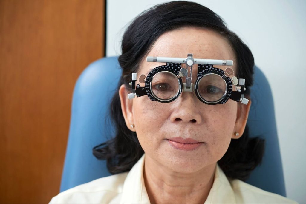
It typically includes a range of tests designed to check various aspects such as visual acuity, eye pressure, retina health, lens clarity, and eye muscle coordination. From this, it’s clear to see that the purpose of an ophthalmic exam is not only to determine your prescription for glasses but also to identify any potential eye diseases or conditions, many of which might not present immediate symptoms.
This makes regular ophthalmic exams crucial for early detection and management of eye health issues, ensuring long-term vision health and quality of life.
What is the Difference Between an Eye Test and an Eye Exam?
Understanding the distinction between an eye test and an eye exam is crucial in comprehending the scope of eye health assessments.
A sight test fundamentally evaluates how well you can see. It measures your visual acuity and determines if you need a prescription for corrective lenses. This type of test might also include additional assessments, such as checking your visual fields and eye pressure. However, a sight test has its limitations and may not always uncover underlying eye conditions that require a more in-depth analysis.
Conversely, as mentioned, an eye examination is an all-encompassing series of tests conducted by an ophthalmologist or an optometrist. It goes beyond basic vision checks to thoroughly assess your ability to focus on and discern objects. Moreover, it includes various other examinations pertaining to the health of your eyes.
What Happens During an Eye Exam?
An eye exam begins with a basic external examination of the eyes, including the eyelids and the surrounding areas. This preliminary check helps in identifying any visible abnormalities or signs of conditions affecting the outer structure of the eye.
The ophthalmologist then proceeds to examine various internal parts of the eye, such as the conjunctiva, sclera, cornea, and iris. Each of these components is carefully inspected for signs of disease or damage. Various tools and equipment may be leveraged for an in-depth assessment, allowing the eye care professional to observe the details of your eye’s structure and function.
Throughout the eye examination process, the focus is not only on identifying vision problems but also on detecting any early signs of eye diseases, many of which can be effectively managed if caught early.
What Does a Comprehensive Eye Exam Typically Include?
As mentioned, a comprehensive eye exam is an extensive evaluation of your eye health and vision. Beyond the standard process of taking note of patient and family health history, it typically encompasses several key elements. This may include, but is not limited to:
- Vision Test: This fundamental part of the exam assesses how well you can see, both with and without any corrective eyewear you may use. It helps determine your prescription and identify any changes in your vision.
- Pupil Reflex Assessment: This test evaluates the response of your pupils to light, which can indicate the health of your optic nerve and overall neurological function.
- Eye Muscle Function Test: This involves checking the muscles controlling eye movements to ensure they are working correctly and in harmony.
- Peripheral Vision Test: Also known as a visual field test, this assesses your ability to see on the sides while focusing straight ahead, helping to detect issues with ‘side’ vision.
- Slit Lamp Examination: Using an upright microscope known as a slit lamp, the eye doctor examines the front parts of your eye, including the cornea, iris, and lens, to check for any abnormalities or diseases.
- Eye Pressure Test: This test measures the pressure inside your eyes, which is essential for detecting glaucoma and other eye conditions related to intraocular pressure.
- Examination of the Back of the Eye: This involves looking at the retina, optic disc, and blood vessels at the back of your eye, providing vital information about conditions like retinopathy, macular degeneration, and more.
Collectively, these tests provide vital information about your eye health and may lead to further discussions on potential treatments, vision correction options, or any necessary actions based on the results.
1. Visual Acuity Test

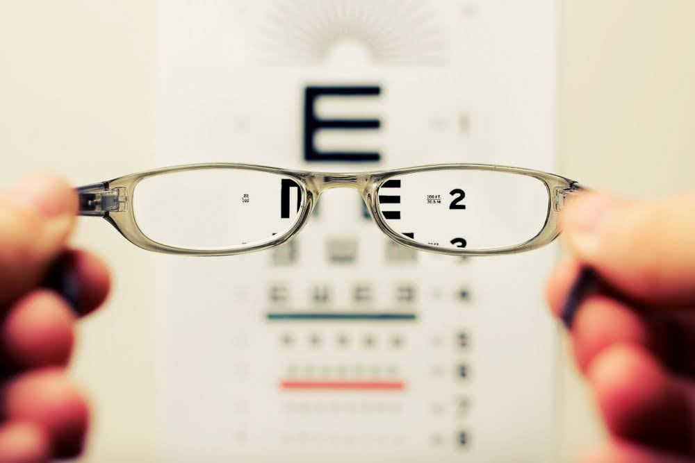
The Visual Acuity Test, commonly known as the “eye chart” test, is a fundamental component of any eye examination. This test typically involves using a Snellen Eye Chart, which displays a series of letters that decrease in size line by line. Positioned approximately 6 metres (20 feet) away from the chart, you will be asked to cover one eye and read the letters aloud, then repeat the process with the other eye. This method assesses how well each eye can see and helps in detecting differences in vision between your eyes.
Additionally, the Near Vision Test is an integral part of assessing visual acuity, especially for tasks performed at a closer range, like reading or using a mobile phone. It involves reading smaller texts at a close distance, and the results will determine how well you can see objects that are near.
What is a Normal Visual Acuity Test Result?
Visual acuity is quantified using a fractional notation, providing valuable information about your eyesight.
In this notation:
- The numerator (the number on top) represents the distance at which you stand from the eye chart, often 20 feet.
- The denominator (the number at the bottom) signifies the distance at which a person with normal vision would be able to read the same line of the chart that you correctly read.
For instance, a measurement of 20/20 means that the line of letters you correctly identified at 20 feet can also be read by an individual with typical vision from the same distance.
A normal visual acuity test result is often denoted as 20/20 vision. However, it’s important to note that 20/20 does not mean perfect vision; it simply indicates that you can see at that specified distance what the average person can see at that distance. Results different from 20/20 may suggest the need for corrective lenses or further eye health evaluation.
2. Colour Vision Test
The Colour Vision Test is another crucial part of an eye examination, specifically designed to screen for colour blindness and assess your ability to distinguish and perceive colours accurately. During this test, you are usually presented with images composed of multicoloured circles, known as Ishihara plates. These images contain numbers or letters formed by dots of different colours and sizes.
The task involves identifying these numbers or letters. The test is effective in detecting various types of colour vision deficiencies, including the most common forms of red-green colour blindness.
What is a Normal Colour Vision Test Result?
A normal colour vision test result is typically determined by the ability to read the majority of the Ishihara plates accurately. Generally, if an individual can correctly identify the images or numbers on ten or more of the first 11 plates, their colour vision is considered normal. Conversely, if a person can correctly read only seven or fewer of these plates, it suggests a deficiency in colour vision.
Do note, however, that there are various other instances where additional tests, including the anomaloscope, might be employed for a more comprehensive assessment of colour vision capabilities.
3. Visual Field Test
The Visual Field Test, also known as the peripheral vision test, assesses the full scope of your vision, including the central and peripheral (side) areas. This test is essential for determining how much you can see without moving your eyes.
Various methods are used for this test, but the most common one is known as the Static Automated Perimetry, which involves an instrument known as an autoperimeter. During the test, you look into the dome-shaped machine while a computer programme flashes small lights at different locations and intensities. You are typically asked to press a button whenever you see these lights. The responses are then recorded to map out your visual field. The results of this test are key in identifying any blind spots or areas of diminished vision, which can be indicators of eye conditions like glaucoma or problems stemming from the optic nerve or brain.
What is a Normal Visual Field Test Result?
Normal visual field test results are represented through various charts that depict the sensitivity and scope of your field of vision. For instance, on the Gray-Scale Map, darker areas highlight parts of the visual field where there may be low sensitivity and, therefore, decreased vision.
Take Action for Your Eye Health with an Eye Examination
Understanding your eye health is essential, but it is equally important to take proactive steps to preserve it. Therefore, regular eye examinations are crucial in early detection and management of potential eye problems. Whether you are experiencing changes in your vision, need an update on your prescription, or simply want to ensure that your eyes are in good health, scheduling an eye exam at EyeWise Vision Clinic is the best course of action.
Don’t wait for symptoms to arise before seeking professional help. Early detection can be key to preventing and treating eye diseases effectively.

Dr. Christopher Khng, specializes in Complex Cataract and Anterior Segment Reconstruction Surgery, in particular, Iris Reconstruction and surgery for Aniridia. His other areas of expertise include Complex Lens surgery, New Lens and Phacoemulsification technologies, Refractive surgery, Phakic IOLs (the Implantable Collamer Lens, ICL), and small-incision, topical anesthesia phacoemulsification cataract surgery. Dr. Khng is a member of the Singapore Medical Association (SMA), a Fellow of the American Academy of Ophthalmology, the American Society of Cataract and Refractive Surgery (ASCRS), and the European Society of Cataract and Refractive Surgery (ECSRS). He is registered with the Singapore Medical Council (SMC) in Singapore and with the General Medical Council (GMC) for practice in the United Kingdom.

