Retinal Detachment
A Guide to Retinal Detachment Symptoms, Treatment, Surgery and Recovery

Dr. Christopher Khng
- Medical Director, Senior Consultant Ophthalmologist
- MBBS, M.Med(Ophth), FRCS(Edin), AMS(Ophth 2003)
Table of Contents
- Retinal Detachment Treatment in Singapore
- Is Retinal Detachment an Emergency?
- What Causes Retinal Detachment?
- Types of Retinal Detachment
- Warning Signs & Symptoms
- Treatment Options
- Macula-On vs Macula-Off: Why Timing Matters
- Recovery After Surgery
- Medisave & Insurance Coverage
- Concerned About Retinal Detachment?
- The Specialist Leading Your Care
- Frequently Asked Questions
- References
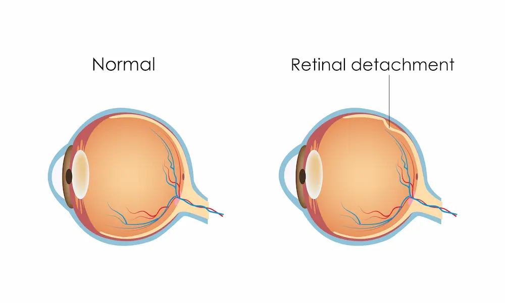
Retinal detachment is a serious eye emergency that requires urgent medical attention. It occurs when the retina, the light-sensitive layer at the back of the eye, separates from its normal position. If left untreated, permanent vision loss can occur.
At EyeWise Vision, patients with retinal tears and retinal detachments receive prompt assessment and treatment. Because retinal detachment can progress quickly, early diagnosis and timely intervention are often critical in preserving vision.
Seek Urgent Assessment
If you are experiencing sudden flashes of light, new floaters, blurred vision or a shadow moving across your vision, seek urgent assessment as soon as possible. Early treatment often leads to better visual outcomes.
Overview
Retinal detachment is a serious eye condition that occurs when the retina separates from the back of the eye. Without prompt treatment, it can lead to permanent vision loss. Common warning signs include sudden flashes of light, new floaters, blurred vision or a dark curtain moving across your field of vision. Treatment may include laser therapy, vitrectomy, scleral buckling or other procedures depending on the type and severity of the detachment.
The specialist leading your care
Our Eye Surgeon

Dr Christopher Khng
Medical Director, Senior Consultant OphthalmologistMBBS, M.Med(Ophth), FRCS(Edin), AMS(Ophth 2003)
Dr Christopher Khng is a Senior Consultant Ophthalmologist with extensive experience diagnosing and treating retinal diseases, including retinal detachment, retinal tears, diabetic retinopathy, and macular disorders.
Dr Khng began his medical education at the University of Aberdeen, Scotland, where he received several academic distinctions, including the Leslie Durno Prize, Alexander Smith Cardno Prize, and Munday and Venn Prize in Medicine. He subsequently completed his medical degree at the National University of Singapore (NUS).
Prior to private practice, he served at leading eye institutions including the Singapore National Eye Centre (SNEC) and Tan Tock Seng Hospital. Drawing on years of specialist training and surgical experience, he provides comprehensive care for patients with retinal conditions, guiding them through every stage of diagnosis, treatment, and recovery.
Dr Christopher Khng
Medical Director, Senior Consultant Ophthalmologist MBBS, M.Med(Ophth), FRCS(Edin), AMS(Ophth 2003)
Is Retinal Detachment an Emergency? Recognising the Warning Signs
Retinal detachment is usually painless, but it often causes sudden changes in vision that should never be ignored.
Sudden Increase in Floaters
Flashes of Light
Dark Shadow or Curtain
Sudden Blurring of Vision
Loss of Peripheral Vision
These symptoms do not always mean a retinal detachment has occurred, but they may indicate a retinal tear or another serious retinal condition that requires urgent evaluation.
When Should You Seek Immediate Treatment?
If you notice a shadow, curtain or veil moving across your vision, do not wait for symptoms to improve.
This may mean the retina is beginning to detach, or that the macula, the part of the retina responsible for detailed central vision, is at risk. Prompt treatment can significantly improve the chances of preserving vision.
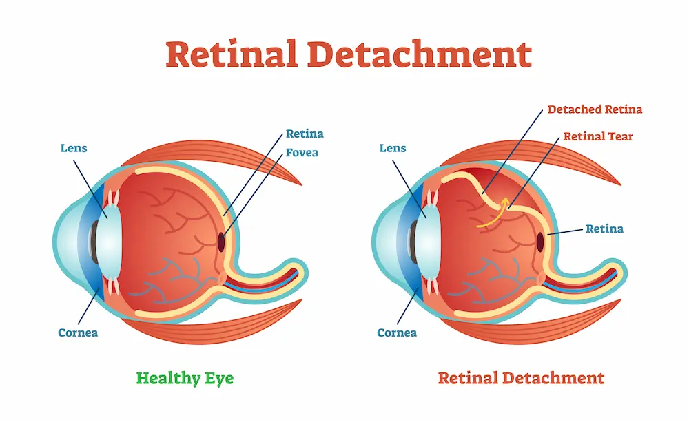
What Causes Retinal Detachment?
Retinal detachment occurs when the retina is pulled away from the back of the eye. There are three main types, each with different causes.
| Type | Mechanism | Common Cause |
|---|---|---|
| Rhegmatogenous | Tear or hole in the retina allows fluid to pass underneat | Most common type. Often related to ageing vitreous changes or high myopia. |
| Tractional | Scar tissue on the retinal surface pulls the retina away | Most commonly associated with advanced diabetic retinopathy. |
| Exudative | Fluid collects beneath the retina without a tear | May be caused by inflammation, vascular conditions or tumours affecting the eye. |
Certain factors can increase the risk of retinal detachment:
- High myopia (severe short-sightedness)
- Previous cataract or eye surgery
- Family history of retinal detachment
- Eye injury or trauma
- Diabetes
- Age-related changes within the eye
- Retinal conditions such as lattice degeneration
Singapore has one of the highest rates of myopia in the world, making regular retinal examinations particularly important for highly myopic individuals.
Types of Retinal Detachment
Understanding the type of retinal detachment helps determine the most appropriate treatment.
- Rhegmatogenous retinal detachment is the most common type and is usually caused by a retinal tear.
- Tractional retinal detachment occurs when scar tissue pulls on the retina, most commonly in patients with diabetic eye disease.
- Exudative retinal detachment develops when fluid accumulates beneath the retina without a tear and is often related to inflammation or other underlying eye conditions.
A detailed retinal examination allows your specialist to identify the type of detachment and recommend the most suitable treatment.
What Happens After a Retinal Detachment Is Diagnosed?
Once retinal detachment is diagnosed, the next step is determining the type and extent of the detachment, as well as whether the macula is involved.
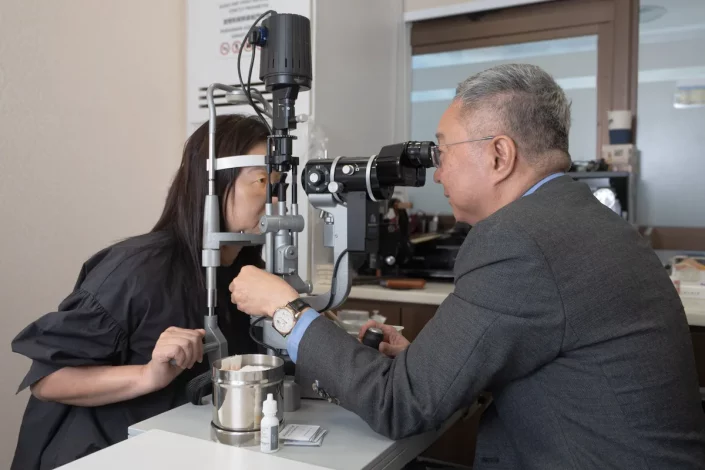
Your retinal specialist will perform a detailed examination and may recommend additional imaging such as OCT scans or retinal photography to better understand the condition and plan treatment.
Depending on the severity of the detachment, treatment may involve laser therapy, vitrectomy, scleral buckling or another appropriate procedure. The recommended treatment will be explained in detail, together with the expected recovery process and follow-up requirements.
After treatment, regular follow-up visits are important to ensure the retina remains attached and is healing as expected.
Treatment Options for Retinal Detachment
Treatment depends on the location and extent of the detachment, the presence of retinal tears and the overall health of the eye.
| Procedure | How it works | Best Suited For | Setting |
|---|---|---|---|
| Laser Photocoagulation | Laser creates a seal around the retinal tear | Retinal tear detected before full detachment | Outpatient |
| Cryotherapy | Controlled freezing seals the retinal tear | Retinal tear detected before full detachment | Outpatient |
| Vitrectomy | Vitreous gel removed; retina repositioned; gas bubble or silicone oil placed to support healing | Most retinal detachments; complex cases | Operating theatre |
| Scleral Buckling | Silicone band placed around the outside of the eye to reduce traction on the retina | Selected cases; often combined with laser or cryotherapy | Operating theatre |
| Pneumatic Retinopexy | Gas bubble injected into eye; positioned against tear; laser or cryotherapy then seals the tear | Selected retinal detachments; less invasive option | Clinic or outpatient |
Vitrectomy
Vitrectomy is one of the most commonly performed procedures for retinal detachment. During the procedure, tiny instruments are used to remove the vitreous gel inside the eye.
This allows the surgeon to repair retinal tears and reposition the retina. A gas bubble or silicone oil may then be placed inside the eye to support the retina while it heals.
Recovery is gradual, and some patients may need to maintain a specific head position for a period after surgery.
Scleral buckling
Scleral buckling involves placing a silicone band around the outside of the eye. This gently indents the wall of the eye and reduces the traction pulling on the retina. The procedure is often combined with laser treatment or cryotherapy and remains an effective treatment option in selected cases.
Pneumatic retinopexy
Pneumatic retinopexy is a less invasive treatment suitable for certain retinal detachments. A gas bubble is injected into the eye and positioned against the retinal tear. Laser treatment or cryotherapy is then used to seal the tear. Careful head positioning is required after the procedure to achieve the best result.
Our team can help you understand which treatment is most appropriate for your condition.
Macula-On vs Macula-Off: Why Timing Matters
One of the most important factors affecting visual recovery is whether the macula remains attached at the time of treatment.
The macula is responsible for the detailed central vision used for reading, driving and recognising faces.
| Status | What it means | Visual Prognosis |
|---|---|---|
| Macula-On | Macula still attached at the time of treatment | Urgent treatment can often preserve excellent vision |
| Macula-Off | Macula has detached before treatment | Surgery can usually reattach the retina, but visual recovery may be less complete |
This is why early diagnosis and treatment are so important.
Recovery After Retinal Detachment Surgery

Recovery varies depending on the type of treatment performed and the severity of the detachment.
In the first few days after surgery, vision is often blurred and mild discomfort is common. If a gas bubble is used, vision will gradually improve as the bubble is absorbed.
Most patients notice progressive improvement over the first few weeks, although complete visual recovery may take several months. Follow-up appointments allow your surgeon to monitor healing and address any concerns during recovery.
If you have been advised to maintain a specific head position after surgery, it is important to follow these instructions carefully to support healing and improve the chances of success.
Medisave & Insurance Coverage for Retinal Detachment Treatment
Retinal detachment treatment is generally claimable under Medisave and most Integrated Shield Plans, subject to policy terms and prevailing regulations.
Our team can assist with insurance pre-authorisation, documentation and estimates of expected out-of-pocket expenses before treatment.
Patients with employer-sponsored medical insurance may also have coverage for retinal surgery and related treatment. We recommend checking your policy details or speaking with our team for assistance.
| Scheme | Coverage | Notes |
|---|---|---|
| Medisave | Eligible inpatient surgical procedures | Available to Singapore Citizens and Permanent Residents, subject to MOH regulations and withdrawal limits |
| Integrated Shield Plans (ISPs) | Private hospital retinal surgery | Coverage depends on policy type, rider and pre-authorisation. Our team can assist. |
| Employer Medical Insurance | May cover retinal surgery and related treatment | Check policy details with your insurer or speak with our team |
Concerned About Retinal Detachment?
Prompt assessment is important when symptoms such as flashes of light, new floaters, blurred vision, or a shadow in your vision develop. Early treatment can often improve the chances of preserving vision and preventing further complications.
If you have concerns about retinal detachment or would like a specialist opinion, arrange a consultation with Dr Christopher Khng.
Early assessment can make a significant difference to your visual outcome.
Frequently asked questions
Retinal Detachment - Patient Information
Can retinal detachment be treated without surgery?
If a retinal tear is detected before detachment occurs, laser treatment or cryotherapy may be sufficient. Once the retina has detached, surgery is usually required.
What is the appropriate treatment for retinal detachment?
The most appropriate treatment depends on the type and severity of the detachment. Options may include laser treatment, vitrectomy, scleral buckling or pneumatic retinopexy. Dr Khng will recommend the most suitable approach after a detailed examination.
How long does recovery take after retinal detachment surgery?
Most patients recover gradually over several weeks to months. The exact timeline depends on the severity of the detachment and the treatment performed.
Can retinal detachment lead to blindness?
Can I fly after retinal detachment surgery?
If a gas bubble has been placed inside the eye, flying is not permitted until the bubble has fully resolved. Changes in air pressure can cause the bubble to expand and damage the eye. Dr Khng will advise when it is safe to travel.
What should I do if I suddenly see flashes or floaters?
While flashes and floaters do not always indicate retinal detachment, they can be warning signs of a retinal tear or other serious retinal condition. A prompt eye examination is recommended. Do not wait for symptoms to worsen before seeking assessment.
Will my vision return to normal after retinal detachment surgery?
Retinal detachment is a sight-threatening condition. Do not delay seeking urgent assessment.
References
- Gariano RF, Kim CH. Evaluation and management of suspected retinal detachment. American Family Physician. 2004;69(7):1691–1698.
- Mitry D, et al. The epidemiology of rhegmatogenous retinal detachment: geographical variation and clinical associations. British Journal of Ophthalmology. 2010;94(6):678–684.
- Heimann H, et al. Scleral buckling versus primary vitrectomy in rhegmatogenous retinal detachment. Ophthalmology. 2007;114(12):2142–2154.
- Ling R, et al. An analysis of the risk of redetachment following retinal reattachment surgery. British Journal of Ophthalmology. 2004;88(12):1522–1524.
- Singapore National Eye Centre. Retinal Detachment. Available at: snec.com.sg [Accessed June 2026].

Contact Us
Have concerns about your eye health or changes in your vision? Our team is here to help. Simply complete the form below, or reach us directly by phone (+65 6476 1211) or WhatsApp for assistance with your appointment.